

Advanced imaging services to support the evaluation of brain, spine, nerve and vascular conditions, helping doctors make informed clinical decisions.
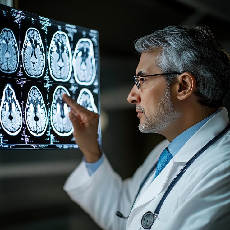
Diagnostic neuroradiology uses specialised imaging techniques to assess the brain, spine, nerves, head and neck. These investigations help clinicians evaluate neurological conditions, identify structural changes and plan appropriate care.
Imaging plays an important role in neurological assessment by providing detailed information about structures that may not be examined directly during a clinical evaluation. The choice of investigation depends on the patient’s symptoms, medical history and the clinical question being addressed.
MRI: Brain, spine and other imaging, including selected advanced MRI techniques.
CT Scan: Detailed imaging of the brain, spine and other structures.
Diagnostic Angiography: Imaging to assess blood vessels when clinically indicated.
Colour Doppler: Ultrasound-based assessment of blood flow.
Computed Radiography (CR): Digital X-ray imaging.
PACS: A system for storing and sharing medical images with clinicians.
The department’s webpage lists a 3 Tesla digital MRI system, multidetector CT scanner, Colour Doppler systems, computed radiography and PACS.
Advanced MRI techniques may include:
These techniques can provide additional information about brain structure, blood flow, tissue characteristics or vascular anatomy in selected cases. Availability and suitability depend on clinical indication and the imaging protocol.
Diagnostic imaging can support the evaluation of neurological symptoms, identify structural or vascular changes and help doctors plan further investigations or treatment.
Imaging may be considered when assessing symptoms such as persistent headaches, seizures, weakness, numbness, changes in coordination or other neurological concerns. However, not every symptom requires imaging. The treating doctor determines whether a scan is appropriate based on the clinical assessment.
Diagnostic neuroradiology plays an important role in evaluating conditions that affect the brain, spine and nervous system. Imaging findings help doctors understand changes in brain and spinal structures, assess blood vessels and investigate symptoms that may require further medical attention.
The imaging results are considered alongside a patient’s symptoms, medical history and clinical examination. This helps the treating specialist determine whether further investigation, monitoring or treatment may be required.
Different imaging techniques provide different types of information about neurological structures. MRI can produce detailed images of the brain and spine, while CT may be used to assess certain conditions that require rapid or detailed structural imaging. Vascular imaging techniques can help evaluate blood vessels when clinically indicated.
Advanced MRI methods, such as diffusion imaging, perfusion imaging, functional MRI and MR spectroscopy, may provide additional information about selected neurological conditions. The choice of imaging technique depends on the clinical question and the protocol recommended by the treating doctor.
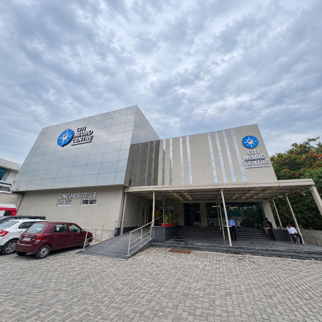
Banjara hills

Trimulgherry

Uppal
Speak with your doctor about the imaging investigation suited to your symptoms and clinical needs. Contact Citi Neuro Centre for appointment and scan-related information.
It is the use of imaging to evaluate the brain, spine, nerves, head and neck.
MRI, CT, angiography, Doppler ultrasound and X-rays may be used depending on clinical need.
Some implants or medical devices require special screening. Inform the imaging team about implants, pregnancy or relevant medical concerns.
Requirements may vary by test and clinical situation. Follow your doctor’s advice and contact the centre for details.
Preparation depends on the scan. Follow instructions from your doctor or imaging team, including any contrast or fasting guidance.